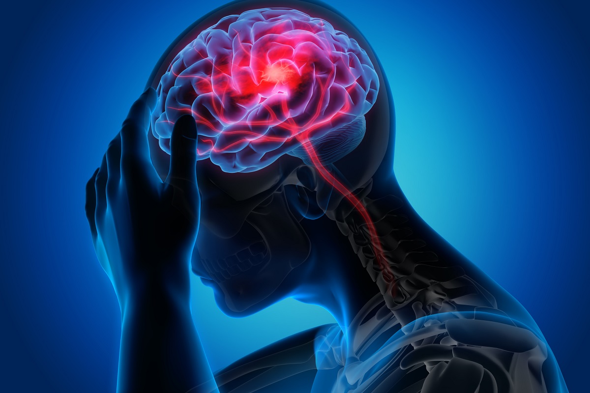
A stroke does not usually announce itself.
There may be no warning. A person may be having breakfast, speaking to the family, walking to the bathroom or getting ready for work—and suddenly the speech becomes unclear, one side of the face droops, an arm becomes weak or the person is unable to walk properly.
The most dangerous response at that moment is often not panic. It is waiting.
Families frequently spend precious minutes trying to understand what has happened: “Maybe BP badh gaya hai.” “Sugar check karo.” “Thoda rest karne do.” “Dekhte hain, theek ho jayega.”
In stroke, these minutes can matter enormously.
A stroke is essentially a brain emergency. In an ischaemic stroke, a blood vessel supplying the brain becomes blocked. In a haemorrhagic stroke, a blood vessel ruptures and bleeding occurs in or around the brain. The treatments are completely different—which is precisely why a family should not try to determine the type of stroke at home.
The first job is simple: Recognise it. Record the time. Get emergency medical help.
THE FIRST QUESTION IS NOT “WHAT IS THE CAUSE?”
It is: “Could this be a stroke?”
The classic warning signs remain remarkably simple. Remember B.E. F.A.S.T.
- B – Balance
Sudden loss of balance, difficulty walking, dizziness or inability to coordinate movements. - E – Eyes
Sudden loss or blurring of vision, double vision or loss of vision in one or both eyes. - F – Face
Ask the person to smile. Does one side of the face droop or feel numb? - A – Arm
Ask the person to raise both arms. Does one arm become weak, numb or drift downward? - S – Speech
Is speech slurred? Is the person unable to speak properly, find words or understand what is being said? - T – Time
Do not wait to see whether it improves. Activate emergency medical services immediately.
The American Stroke Association specifically recommends B.E. F.A.S.T. recognition, while the CDC emphasises sudden weakness or numbness, speech or comprehension difficulty, visual disturbance, imbalance and sudden severe headache as important warning signs.
There is one additional point families should remember: A stroke does not always look like paralysis.
Sudden confusion, difficulty understanding speech, isolated visual symptoms, severe imbalance or a sudden unexplained neurological change can also represent stroke.
And sometimes the symptoms disappear. That does not make the event harmless.
A transient ischaemic attack—or TIA—is a warning event in which stroke-like symptoms resolve. At home, it may be impossible to know whether it was a TIA or an evolving stroke. It still requires urgent medical assessment.
FIRST AID FOR STROKE: WHAT SHOULD THE FAMILY ACTUALLY DO?
When stroke is suspected, the family has a very important role—but it is not to treat the stroke at home. It is to prevent delay.
DO THESE FIVE THINGS
1. Note the exact time.
This is one of the most valuable pieces of information you can give the stroke team.
If the patient was last seen completely normal at 8:00 AM but symptoms were discovered at
9:30 AM, tell the medical team exactly that.
If the patient wakes up with symptoms, note the last time they were known to be normal,
usually before going to sleep.
The treatment decision may depend on this timeline.
2. Call emergency medical services and arrange rapid transport.
Do not spend 30–40 minutes calling multiple relatives, neighbours or doctors before
arranging emergency transport.
The ambulance journey is part of the treatment pathway. Emergency personnel can begin
assessment, communicate with the receiving hospital and help expedite care. The CDC
specifically advises against driving the patient yourself when emergency medical transport
is available.
3. Keep the patient safe and under observation.
If the patient is conscious, keep them comfortable and supervised.
If they are drowsy or have reduced consciousness, protect the airway and position them
safely while awaiting professional help.
If they stop breathing normally, emergency resuscitation measures may be required.
4. Carry the important information.
Take, if readily available:
Current medication list
Details of blood thinners or antiplatelet medicines
Previous stroke history
Diabetes and hypertension history
Recent medical records
Previous brain scans, if easily accessible
Information about when the patient was last known to be normal
But do not delay transport while searching for old reports.
5. If you have a glucometer and know how to use it, checking blood glucose can be
useful—but it must not delay emergency activation.
Low blood sugar can mimic some stroke symptoms. The 2024 AHA/American Red Cross first-aid
guidance considers glucose measurement reasonable for trained/equipped first-aid providers
when it does not delay emergency activation.
SOME OF THE MOST COMMON FAMILY MISTAKES, I WISH WE COULD AVOID
This is where awareness can genuinely save brain function.
Do not give aspirin on your own.
This is one of the most common misconceptions. People often assume: “Stroke hua hai, aspirin de do.” But not every stroke is caused by a clot.
A haemorrhagic stroke is caused by bleeding. Giving aspirin when there is intracranial bleeding can potentially worsen the situation.
The American Stroke Association explicitly advises that aspirin should not be taken during an acute stroke before the stroke type is established, because some strokes are caused by ruptured blood vessels.
The correct sequence is diagnosis first, treatment second.
Do not give food, water, juice or medicines by mouth if the person's swallowing is impaired, consciousness is reduced or you are unsure whether they can swallow safely.
Stroke can affect swallowing. A patient who looks awake may still have significant dysphagia and can aspirate food or liquid into the lungs. Dysphagia screening is therefore an important component of acute stroke care.
So please do not force: Water Tea or coffee Juice Food Tablets Ayurvedic or herbal preparations into the mouth of a person with suspected stroke.
Do not massage the paralysed arm or leg.
Massage does not reopen a blocked cerebral artery. Nor does vigorously moving the affected limb treat the stroke. The priority is emergency evaluation and reperfusion where appropriate.
Do not wait for BP or sugar to “come down” before seeking help.
Checking blood pressure can be useful. But repeatedly checking it while delaying hospitalisation is not.
Similarly, a high blood pressure reading during an acute stroke does not automatically mean that the family should give an extra dose of antihypertensive medication. Acute blood pressure management in stroke is nuanced and depends on the stroke type and whether the patient is being considered for reperfusion therapy.
Do not assume that improvement means the emergency is over.
A patient whose speech becomes normal after 10 minutes may still have experienced a TIA or a transient stroke syndrome. The symptoms disappearing is not the same as the risk disappearing.
Let's first find the best hospital.
Choosing an appropriate stroke-capable facility matters, but searching endlessly for the “perfect” hospital can itself become dangerous.
The 2026 guideline emphasises organised stroke systems and, depending on the local system, consideration of direct transport to an endovascular-thrombectomy-capable centre where rapid interhospital transfer is not reliably available.
The best hospital is the one that can deliver the appropriate treatment quickly and safely.
THE STROKE TREATMENT LANDSCAPE HAS CHANGED
For many years, families heard a simple message: “Stroke treatment is possible if the patient reaches within 4.5 hours.” That message was important—but modern stroke medicine is more sophisticated.
The 2026 American Heart Association/American Stroke Association guideline has replaced the 2018 guideline and its 2019 update and incorporates major advances in intravenous thrombolysis, endovascular thrombectomy, imaging, prehospital care and patient selection.
1. Tenecteplase has become an important thrombolysis option
For eligible adults with acute ischaemic stroke within the conventional 4.5-hour window, the 2026 guideline recommends either tenecteplase or alteplase for intravenous thrombolysis.
This does not mean that every stroke patient receives thrombolysis. The patient must first be assessed appropriately, brain imaging must exclude contraindications such as intracranial haemorrhage, and the neurological deficit and clinical circumstances must be evaluated.
The important message for families is: Do not decide at home that the patient is “too old”, “too mild”, or “too late” for treatment. Let a stroke team make that decision.
2. The window for treatment is not always limited to 4.5 hours
Selected patients who wake up with stroke symptoms or present beyond the conventional thrombolysis window may still be considered for intravenous thrombolysis when advanced imaging demonstrates salvageable brain tissue.
The 2026 guideline allows consideration of IV thrombolysis in selected patients approximately 4.5–9 hours from last known well, or in certain wake-up strokes, when appropriate perfusion imaging identifies salvageable tissue.
This is one reason why the family should never decide that treatment is impossible simply because several hours have passed.
MECHANICAL THROMBECTOMY: WHEN A CLOT CAN BE PHYSICALLY REMOVED
One of the biggest advances in modern stroke care has been mechanical thrombectomy.
In selected patients with a large-vessel occlusion, an endovascular specialist can navigate a catheter through the blood vessels to reach the blocked artery and remove the clot.
The 2026 guidelines have further expanded the population that may benefit.
For selected patients with anterior-circulation large-vessel occlusion, thrombectomy can be recommended within the 6-hour window, and selected patients may remain candidates in the 6–24-hour window based on clinical and imaging criteria. Importantly, newer evidence has also expanded treatment to some patients with a larger area of established brain injury than would previously have been considered suitable.
The guideline also provides a strong recommendation for selected patients with basilar artery occlusion within 24 hours under specified clinical and imaging criteria.
This is a major change in the way we think about stroke. The question is no longer simply: “How many hours have passed?” It is increasingly: “How much brain is already irreversibly injured—and how much potentially salvageable brain remains?” That is why rapid imaging and expert interpretation matter.
“TIME IS BRAIN” — BUT TIME IS NOT THE ONLY THING THAT MATTERS
The phrase “time is brain” remains absolutely relevant. But modern stroke care has added another dimension: “Time + Tissue + Technology + Team.”
A patient may reach the hospital several hours after symptom onset and still have potentially treatable brain tissue. Another patient arriving earlier may already have a large established infarction.
Therefore, modern stroke treatment combines: Clinical assessment Brain imaging Vascular imaging when appropriate Assessment of the severity and disability caused by the stroke Identification of large-vessel occlusion Evaluation of salvageable brain tissue where indicated Rapid decision-making by a coordinated stroke team
The 2026 guideline also emphasises that additional imaging should not create unnecessary delays in patients who are already eligible for rapid treatment.
STROKE IS NOT TREATED BY ONE DOCTOR — IT IS TREATED BY A TEAM
A modern stroke unit is not simply a neurologist sitting in an emergency room. Stroke care is a coordinated chain in which every minute, every decision and every handover matters.
At the Institute of Neurosciences, Medanta Noida, the approach to acute stroke brings together neurology, neurointervention, emergency medicine, neurocritical care, neuroradiology/imaging, neurosurgery, anaesthesia and rehabilitation when required.
The neurologist is often the first person to establish that the patient's symptoms could represent an acute stroke, assess the neurological deficit and determine the immediate treatment pathway. But if the imaging shows a large-vessel blockage that may require thrombectomy, the next step may involve the neurointervention team. If the patient is critically ill, neurocritical-care expertise becomes essential. And once the immediate emergency has been addressed, rehabilitation becomes an integral part of the patient's recovery.
At Medanta Noida, the neurovascular service includes expertise in acute ischaemic stroke, mechanical thrombectomy and other neurovascular disorders, supported by an advanced endovascular setup including an Icono biplane cath lab.
The important point is that these are not independent services working in isolation. The patient moves through a pathway. The team moves with the patient. That distinction matters enormously in stroke.
A patient may arrive with facial asymmetry and weakness. The neurologist evaluates the clinical picture. Brain imaging establishes whether there is bleeding or evidence of ischaemic injury. Vascular imaging may identify an occluded artery. The neurointervention team can then determine whether the patient is a candidate for thrombectomy. Critical-care specialists may manage airway, blood pressure and other physiological issues in a severely affected patient. Rehabilitation specialists subsequently work on mobility, speech, swallowing and functional recovery.
This is what an integrated stroke-care model should mean: not simply having several specialists in the same hospital, but having the right expertise available at the right point in the patient's journey.
ADVANCED IMAGING: SOMETIMES THE SCAN TELLS US MORE THAN THE CLOCK
One of the most important changes in modern stroke medicine is the way we use imaging.
For many years, the central question was: “How long ago did the stroke start?” Today, we increasingly ask another question: “How much brain has already been irreversibly injured, and how much may still be saved?”
This is where advanced imaging becomes extremely important.
At Medanta Noida, the neuroscience service has access to advanced neuroimaging including 3.0 Tesla MRI and 256-slice CT, while the neurovascular service also uses CT angiographic imaging for evaluation of cerebral and cervical vessels.
In a suspected acute stroke, the initial CT scan can rapidly answer one of the most important questions: Is there bleeding?
If there is intracranial haemorrhage, the treatment pathway is completely different from that of an arterial blockage.
But in suspected ischaemic stroke, imaging can go much further.
CT Angiography (CTA)
CTA can show whether a major cerebral artery is blocked. This is particularly important when we suspect a large-vessel occlusion (LVO)—the type of blockage that may make a patient eligible for mechanical thrombectomy.
The 2026 AHA/ASA guideline specifically identifies CT angiography or MR angiography as a critical component of emergency imaging and triage when large-vessel occlusion is suspected.
CT Perfusion (CTP) and MR Perfusion
In selected patients, perfusion imaging can provide another layer of information. It helps clinicians estimate: the approximate infarct core—brain tissue that is already irreversibly injured; the surrounding hypoperfused but potentially salvageable tissue, often referred to as the ischaemic penumbra; and the mismatch between the two.
This becomes particularly valuable when the time of onset is uncertain or when the patient presents outside the conventional treatment window.
For example, a person may wake up in the morning with weakness. We may not know exactly when the stroke occurred. Traditionally, uncertainty about the onset time created a major limitation.
Today, appropriately selected patients can sometimes be evaluated using MRI diffusion/FLAIR mismatch or perfusion imaging to determine whether there is potentially treatable tissue. The 2026 AHA/ASA guideline incorporates DWI-FLAIR and perfusion-based selection strategies for selected extended-window thrombolysis patients.
But there is an important caveat. Advanced imaging should guide treatment—not become a reason to delay treatment.
For patients presenting within the early thrombectomy window, the 2026 guideline notes that advanced tissue imaging such as CT perfusion or MR perfusion is not necessarily required before thrombectomy in every eligible patient. Additional imaging should not unnecessarily exclude patients who may benefit or delay reperfusion.
That is an important distinction. Technology is valuable only when it accelerates the right decision. The goal is not to perform every possible scan. The goal is to obtain the right information, quickly enough to act on it.
FROM “TIME IS BRAIN” TO “TIME + TISSUE”
This is perhaps the most important message I would like families to understand. We should never interpret the newer imaging technology as permission to wait.
The four-hour or four-and-a-half-hour window is not a waiting period. It is an emergency treatment window.
A patient with suspected stroke should reach medical care as quickly as possible, even if the family thinks that several hours have already passed.
Once the patient reaches a stroke-capable centre, the clinical examination and imaging help us determine what treatment remains possible.
In selected patients, the combination of: clinical assessment + brain imaging + vascular imaging + tissue imaging can identify patients who may still benefit from reperfusion treatment despite an uncertain or extended time of onset.
This is one of the most remarkable developments in stroke medicine. We are increasingly moving from a simple “time-based” model to a more sophisticated “time-and-tissue-based” model.
But the message for the public remains remarkably simple: Never lose time at home because you think the treatment window has already closed. Let the stroke team make that decision.
WHY STROKE AWARENESS MATTERS SO MUCH TO ME
There is something uniquely unforgiving about stroke. With many illnesses, a delay of an hour may change little. With stroke, the same hour can change whether a person walks independently, returns to work, communicates normally or requires long-term assistance.
That is why stroke care cannot begin when the patient enters the hospital. It begins at home.
It begins when a spouse recognises that the smile is asymmetric. It begins when a son notices that his father's speech suddenly sounds different. It begins when a daughter realises that her mother cannot lift one arm.
And it begins when somebody says: “This could be a stroke. Let's not wait.”
THE BIGGER MESSAGE
Stroke treatment has entered an era where more patients can be treated, more patients can be assessed with greater precision, and treatment decisions can be guided by both time and tissue. But advanced treatment can help only when the patient reaches the right system quickly enough. The most sophisticated thrombectomy device cannot compensate for a three-hour delay at home. The most advanced imaging cannot reverse the damage caused by avoidable hesitation.
And no guideline can save a patient who never reaches medical care.
So remember three things: Recognise the signs. Record the time. Reach emergency stroke care immediately.
Because when it comes to stroke, the first treatment is not a tablet. The first treatment is time.
“In modern stroke care, the clock tells us when the patient arrived. Advanced imaging can help us understand what is happening to the brain. The combination of both allows us to make better treatment decisions.”
— Dr. Kapil Kumar Singhal
Director – Neurology, Institute of Neurosciences, Medanta Noida
The Future of Women's Surgical Care
Stroke: When Every Minute Can Change the Brain

Beyond Smoking: How Advanced Diagnostics Are Changing the Future of Lung Cancer Care
The Evolution of Modern Spine Surgery
Beyond Awareness: The Critical Role of Early Cancer Diagnosis
Robotic Surgery: Precision in the Hands of the Surgeon
Behind the Mask: The Human Side of Neurointervention with Dr. Tariq Matin

Womanhood Is a Journey — Health Should Be Part of Every Stage

India's Metabolic Wake-Up Call
Endoscopic Surgery Helps Preserve Vision After Complex Orbital Injury: Dr. Harnarayan Singh
Interventional Radiology: The Life-Changing Treatment You've Probably Never Heard Of!
Kidneys at Work: The Silent Guardians of Your Health
Your Heart May Be Talking. Are You Listening?
Why Are More Young Adults Facing Nerve Damage, Numbness & Back Pain?
Robotic Uro-Oncology Surgery: Precision, Innovation and Better Patient Outcomes
Vape Culture: The Hidden Trap Among Youth

Flu Cases Rise in NCR: Dr. Abhijeet Singh on Symptoms, Treatment & Prevention